

Regenerative medicine continues to redefine the boundaries of modern healthcare, offering innovative solutions for conditions that were once considered irreversible or purely manageable through symptom mitigation. However, the ultimate success of these advanced cellular therapies relies intimately on the meticulous steps taken long before any biological material is extracted or applied. A rigorous Stem Cell Diagnosis and Evaluation process forms the absolute foundation of regenerative care. This crucial preliminary phase ensures that cellular interventions are not only medically appropriate but also highly optimized for the individual patient’s unique physiological landscape. By thoroughly investigating the underlying pathology and the patient’s overall biological profile, medical professionals can accurately determine the potential efficacy of regenerative interventions.
The Imperative of Precise Patient Selection
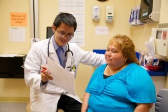
The concept of utilizing the body’s own biological mechanisms to heal damaged tissue is universally appealing, yet it is medically imperative to recognize that cellular therapies are not a universal panacea. Patient selection is arguably the most determinative factor in regenerative medicine outcomes. Physicians must conduct an exhaustive review of the patient’s medical history, meticulously documenting prior injuries, chronic systemic conditions, active infections, and current pharmaceutical regimens. Factors such as advanced age, severe metabolic disorders, or compromised immune systems can significantly influence how the body responds to regenerative treatments. Therefore, a thorough diagnostic workup is essential to filter out candidates for whom the procedure might be ineffective or medically contraindicated, thereby upholding the highest ethical and safety standards.
Advanced Imaging and Structural Mapping
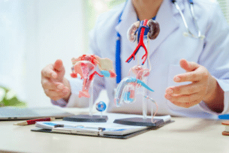
A cornerstone of the pre-treatment assessment involves high-resolution diagnostic imaging. To accurately target cellular therapies, physicians must have a precise, multi-dimensional understanding of the damaged tissue environment. Magnetic Resonance Imaging (MRI) is frequently utilized to evaluate soft tissue structures, including cartilage degradation, ligamentous tears, and precise neurological lesions. X-rays provide necessary baseline data regarding bone density and skeletal alignment, particularly in severe orthopedic cases like advanced osteoarthritis. Furthermore, diagnostic musculoskeletal ultrasound offers real-time dynamic assessment of joint mechanics and tendon integrity. These advanced imaging modalities not only pinpoint the exact location for future cellular delivery but also establish a quantifiable baseline, allowing specialists to objectively measure tissue regeneration and structural improvement following the therapy.
Laboratory Analysis and Systemic Health Profiling
Beyond structural imaging, systemic physiological health plays a massive role in the success of cellular treatments. Extensive laboratory testing is a mandatory component of the evaluation phase. Blood panels are drawn to assess vital organ function, evaluate complete blood counts, and screen for underlying infectious diseases or autoimmune markers that could interfere with cellular engraftment. Assessing the patient’s inflammatory markers is particularly crucial; a highly inflamed systemic environment can prematurely degrade introduced stem cells or push them toward an undesirable lineage. By identifying systemic imbalances during the diagnostic phase, physicians can implement pre-treatment optimization protocols—such as nutritional interventions or temporary anti-inflammatory management—to create a more hospitable biological environment for the incoming regenerative cells.
Assessing Cellular Viability and Reserve


When considering autologous treatments, wherein the biological material is harvested directly from the patient, medical professionals must evaluate the quality and availability of the patient’s own cellular reserves. The regenerative capacity of bone marrow or adipose tissue can vary drastically from one individual to another. Specialized institutions dedicated to international healthcare standards, such as Liv Hospital, employ sophisticated methodologies to estimate cellular yield and viability prior to invasive harvesting. If a patient’s autologous reserves are deemed insufficient due to age or underlying pathology, the medical team can strategically pivot the treatment plan to explore allogeneic (donor-derived) options or adjunctive biological therapies, ensuring no time or resources are wasted on a suboptimal procedure.
Multidisciplinary Review and Baseline Establishment
The most effective diagnostic protocols involve a collaborative, multidisciplinary approach. Complex degenerative conditions often require the combined expertise of orthopedic surgeons, neurologists, hematologists, and interventional radiologists to formulate a cohesive treatment strategy. This collaborative evaluation ensures that every facet of the patient’s condition is considered. During this phase, realistic expectations are established. Patients receive transparent, scientifically grounded data regarding projected outcomes, potential risks, and anticipated recovery timelines. Furthermore, this rigorous diagnostic period establishes the critical objective baseline parameters—ranging from pain scales and mobility metrics to functional capacity tests—that will be utilized to scientifically track the patient’s progress and validate the long-term success of the cellular intervention over the coming months and years.