
Introduction
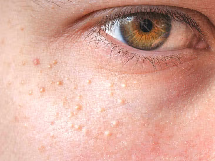
Tiny white or yellowish bumps scattered around the eyes, cheeks, or forehead can be frustrating. These blemishes—known as milia—are keratin-filled cysts that form just beneath the surface of the skin. Although harmless and painless, they often prove stubborn against standard acne remedies and over-the-counter exfoliants. This comprehensive guide explores what causes milia, why some people are more prone than others, and how Milia Removal Treatment can permanently clear the complexion while minimizing scarring and recurrence.
- Introduction
- What Exactly Are Milia?
- Why Do Milia Form?
- When Should You Seek Professional Care?
- In-Clinic Milia Removal Techniques
- 1. Manual Extraction
- 2. Electrocautery
- 3. Radiofrequency (RF) Ablation
- 4. CO₂ or Erbium:YAG Laser
- Pros of Professional Removal
- Home Care: What Works and What Doesn’t
- The Treatment Appointment: Step-by-Step
- Post-Treatment Care and Long-Term Prevention
- Potential Risks and Side Effects
- Conclusion
What Exactly Are Milia?
Milia (singular: milium) are miniature cysts composed of trapped keratin—a structural protein found in skin, hair, and nails. Unlike whiteheads, they have no visible pore or opening, which explains why squeezing rarely works and can even lead to scarring. Dermatologists classify milia into two broad categories:
- Primary Milia– Develop spontaneously when dead skin cells fail to exfoliate naturally.
- Secondary Milia– Form after trauma, burns, blistering disorders, or the prolonged use of topical steroids.
Because the lesions sit under an intact epidermis, they present as firm, dome-shaped papules without redness or inflammation.
Why Do Milia Form?
Multiple factors contribute to their appearance:
- Skin Maturation– Newborns frequently develop primary milia that self-resolve as the skin adapts to life outside the womb.
- Sun Damage– Chronic UV exposure thickens the epidermis, trapping keratin beneath the surface.
- Heavy Cosmetics– Occlusive makeup, sunscreens, or thick moisturizers impede natural exfoliation.
- Genetics & Hormones– Some individuals simply inherit a propensity for sluggish cell turnover.
- Underlying Skin Conditions– Disorders like rosacea or burns can trigger secondary milia through disruption of normal skin architecture.
Understanding these drivers helps tailor both professional and at-home prevention strategies.
When Should You Seek Professional Care?
While single milia occasionally disappear, clusters around delicate areas—especially the under-eye zone—often persist for years. Professional evaluation is warranted when:
- The bumps are numerous, spreading, or cosmetically bothersome.
- Over-the-counter retinoids or exfoliants have failed after six to eight weeks.
- Lesions arise suddenly following trauma, suggesting secondary milia.
- You’re prone to post-inflammatory hyperpigmentation and want to avoid scarring.
Dermatologists not only remove existing cysts but can rule out masquerading conditions such as syringomas or xanthelasma.
In-Clinic Milia Removal Techniques
1. Manual Extraction
The dermatologist cleanses the area, applies a sterile lancet to create a pinpoint opening, and gently lifts out the keratin plug with a comedone extractor. Done correctly, this leaves only a tiny pin-prick mark that heals within days.


2. Electrocautery
Low-level electric current delivers heat through a fine needle, vaporizing the superficial cyst. Electrocautery excels for clustered lesions and has minimal downtime, though post-treatment erythema can last 24–48 hours.
3. Radiofrequency (RF) Ablation
RF energy converts into heat at the tip of a micro-probe, breaking down the cyst while sealing blood vessels. This method offers pinpoint accuracy—ideal for milia around the eyelids—without significant collateral damage.
4. CO₂ or Erbium:YAG Laser
Fractionated or focused lasers ablate the overlying epidermis, exposing the keratin for easy removal. Lasers are particularly useful when milia coexist with photodamage, allowing simultaneous resurfacing and tightening.
Pros of Professional Removal
| Benefit | Why It Matters |
| Minimal Scarring | Tiny incisions heal quickly |
| Immediate Results | Lesions vanish in a single session |
| Lower Recurrence Rate | Complete removal of the cyst lining |
| Sterile Technique | Reduces infection and pigmentation risk |
Home Care: What Works and What Doesn’t
Dermatologists often recommend adjunct measures to reduce new lesions:
- Topical Retinoids– Tretinoin or adapalene accelerates cell turnover.
- Chemical Exfoliation– Low-strength glycolic or salicylic acid frees trapped keratin.
- Lightweight Moisturizers– Look for non-comedogenic labels to avoid pore occlusion.
- Broad-Spectrum SPF– Prevents UV-induced epidermal thickening.
Avoid digging with needles or trying “pimple-popper” gadgets at home; improper technique invites infection, scarring, and post-inflammatory pigmentation, especially on darker skin tones.
The Treatment Appointment: Step-by-Step
- Consultation and Skin Analysis
Your clinician determines whether you have primary or secondary milia and discusses health history, medication use, and skin-care routine. - Cleansing and Preparation
Antiseptic solutions remove surface bacteria. For sensitive patients, a topical anesthetic cream may be applied. - Precise Lesion Breakdown
The selected technique—extraction, electrocautery, RF, or laser—targets each cyst individually, ensuring minimal trauma. - Aftercare Application
An antibiotic ointment or soothing thermal-water gel reduces redness and forms a protective barrier. - Post-Procedure Instructions
You’ll receive guidelines on cleansing, moisturizing, and sun avoidance to speed recovery and prevent recurrence.
Most sessions last 15–30 minutes, and patients can resume normal activities immediately.
Post-Treatment Care and Long-Term Prevention
- Gentle Cleansing– Twice-daily washing with a non-foaming, pH-balanced cleanser prevents residue build-up.
- Moisture Balance– Swap heavy creams for gel-based or ceramide-enriched lotions.
- Weekly Exfoliation– Enzyme masks or low-strength acids remove dead skin cells softly.
- Monitor Sun Exposure– Daily mineral sunscreen plus wide-brimmed hats curb UV-related keratin trapping.
- Regular Follow-Ups– Annual dermatology visits catch any emerging lesions early.
Adhering to these habits keeps pores clear and cell turnover efficient, minimizing the chance that milia reappear.
Potential Risks and Side Effects
Though complications are rare when a qualified practitioner performs the procedure, possible issues include:
- Transient Redness or Swelling– Typically subsides within 48 hours.
- Pigment Alterations– Individuals with Fitzpatrick IV–VI skin types should discuss laser settings to avoid hypopigmentation.
- Micro-scarring– Uncommon, but meticulous aftercare with silicone gels or scar creams can mitigate marks.
Promptly report persistent redness, tenderness, or discharge—signs of infection—to your dermatologist.
Conclusion
Milia may be harmless, but their stubborn nature often calls for professional intervention. From quick manual extraction to sophisticated laser ablation, today’s dermatologic arsenal offers safe, effective solutions tailored to lesion type, location, and skin tone. Combine expert Milia Removal Treatment with smart at-home skincare, and you can look forward to a clearer, smoother complexion—no pinching, scarring, or endless trial-and-error required.