
Blood tests are among the most common diagnostic tools in modern medicine, providing critical insights into everything from cholesterol levels to infection markers. Behind the scenes of every accurate blood test result lies a sophisticated piece of laboratory equipment: the serum separator tube. This specialized collection device plays a crucial role in ensuring that your blood samples are processed correctly, leading to reliable diagnostic results that healthcare providers depend on for treatment decisions.
- What Is a Serum Separator Tube?
- The Gel Barrier Technology
- How Centrifugation Creates Perfect Separation
- Clot Activation and Timing
- Laboratory Processing and Quality Control
- Common Testing Applications
- Advantages Over Traditional Collection Methods
- Potential Limitations and Considerations
- Looking Forward: Innovations in Blood Collection
- Understanding Your Blood Test Results
Understanding how serum separator tubes work reveals the intricate science that makes modern blood testing both accurate and efficient. From the moment blood is drawn to the final laboratory analysis, these tubes facilitate a complex separation process that isolates the liquid portion of blood from its cellular components. This separation is essential for most blood chemistry tests, hormone analyses, and protein measurements that doctors use to assess your health.
Whether you’re a healthcare professional seeking to understand laboratory processes better or a curious patient wondering about the tubes used during your blood draw, exploring the science behind serum separator tubes offers valuable insights into modern diagnostic medicine. The technology represents decades of innovation in laboratory science, combining materials engineering with biological understanding to create tools that consistently deliver accurate results.
What Is a Serum Separator Tube?
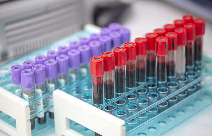
A serum separator tube is a specialized blood collection device designed to separate serum from blood cells through centrifugation. Unlike plasma, which contains clotting factors, serum is the clear, yellowish liquid that remains after blood has clotted and been separated from its cellular components. This distinction is crucial for many laboratory tests that require pure serum samples free from cellular interference.
The tube itself consists of several key components that work together to achieve clean separation. The walls are typically made of glass or plastic treated with silicone to prevent blood cells from sticking. Most importantly, the tube contains a thixotropic gel barrier that moves during centrifugation to create a physical barrier between the serum and the clotted blood cells below.
Modern serum separator tubes also contain clot activators, usually silica particles or glass beads, which accelerate the natural clotting process. This activation reduces the time needed for complete clot formation from the typical 30-60 minutes down to just 15-30 minutes, improving laboratory efficiency while maintaining sample integrity.
The Gel Barrier Technology
The gel barrier represents the most ingenious aspect of serum separator tube design. This specialized material remains viscous at room temperature but becomes fluid when subjected to the centrifugal forces used in laboratory processing. During centrifugation, the gel moves upward through the sample due to its specific density, which falls between that of serum and clotted blood cells.
As the centrifuge spins, the gel forms a stable, impermeable barrier that prevents cellular components from contaminating the serum above. This physical separation is maintained even during subsequent handling and storage, protecting sample integrity for extended periods. The gel’s unique properties ensure that it won’t dissolve into the serum or interfere with most analytical procedures.


Different manufacturers have developed proprietary gel formulations optimized for specific testing requirements. Some gels are designed to be completely inert to avoid interfering with drug level measurements, while others include additives that enhance separation efficiency for challenging samples with high lipid content or unusual cellular distributions.
How Centrifugation Creates Perfect Separation
Centrifugation transforms the mixed blood sample into distinct layers based on density differences between components. When a serum separator tube is placed in a centrifuge and spun at high speeds—typically 1,000 to 1,300 times the force of gravity—the heavier blood cells are forced to the bottom of the tube while the lighter serum rises to the top.
The process begins within minutes of centrifugation as red blood cells, being the densest component, settle at the bottom. White blood cells and platelets form a thin intermediate layer called the buffy coat, while the serum occupies the upper portion of the tube. The gel barrier, activated by the centrifugal force, migrates upward and positions itself between the cellular components and the serum.
This layered separation must occur under precise conditions to ensure optimal results. Temperature, centrifugation speed, and duration all affect separation quality. Too little force may result in incomplete separation, while excessive force can cause hemolysis—the breakdown of red blood cells that releases their contents into the serum, potentially interfering with test results.
Clot Activation and Timing
The clotting process in serum separator tubes is carefully controlled through the use of specific activating agents. Silica particles are the most common activators, providing a surface that triggers the blood’s natural coagulation cascade. These microscopic particles activate Factor XII, initiating a series of biochemical reactions that ultimately convert fibrinogen into fibrin threads, forming the clot structure.
Timing is critical in this process. The blood must be allowed sufficient time to clot completely before centrifugation begins. Premature centrifugation can result in fibrin strands contaminating the serum layer, while excessive clotting time may lead to cellular breakdown and analyte degradation. Most protocols recommend a clotting time of 15-30 minutes at room temperature before processing.
Glass beads serve as an alternative activation method, particularly useful for samples from patients on anticoagulant therapy. These beads provide a larger surface area for activation and can sometimes achieve clotting in samples that might not clot effectively with silica alone. The choice of activator often depends on the specific tests being performed and the patient’s clinical condition.
Laboratory Processing and Quality Control
Laboratory technicians follow strict protocols when handling serum separator tubes to ensure consistent, high-quality results. Upon arrival in the laboratory, tubes are inspected for proper fill levels, clot formation, and any signs of hemolysis or contamination. Tubes that don’t meet quality standards are rejected and new samples requested.
The centrifugation process itself requires precise control of multiple variables. Modern laboratory centrifuges are calibrated regularly to ensure accurate speed and force measurements. Temperature monitoring prevents overheating that could denature proteins or affect analyte stability. After centrifugation, technicians verify that complete separation has occurred and that the gel barrier is properly positioned.
Quality control extends beyond processing to include pre-analytical factors that can affect results. Proper patient preparation, correct tube selection for specific tests, and appropriate storage conditions all contribute to reliable outcomes. Many laboratories use automated systems to track samples from collection through analysis, reducing human error and improving traceability.
Common Testing Applications
Serum separator tubes are essential for a wide range of diagnostic tests that healthcare providers order routinely. Chemistry panels measuring glucose, electrolytes, liver enzymes, and kidney function markers all require serum samples. These tests analyze dissolved substances in the blood that would be diluted or contaminated by the presence of blood cells.
Hormone testing represents another major application for serum separator tubes. Thyroid hormones, reproductive hormones, and stress hormones like cortisol are all measured in serum samples. The clean separation provided by these tubes ensures that cellular enzymes don’t interfere with the delicate immunoassays used for hormone measurement.
Protein analysis, including albumin, total protein, and specific protein markers, also relies on serum separator tubes. These tests help assess nutritional status, liver function, and inflammatory conditions. The absence of cellular proteins in properly separated serum ensures accurate measurement of the proteins that doctors need to evaluate.
Advantages Over Traditional Collection Methods
Before serum separator tubes became standard, laboratories used plain glass tubes that required manual separation of serum from clotted blood. This process was time-consuming and prone to contamination as technicians had to carefully pipette serum from the top of the sample without disturbing the clot below.
The integrated gel barrier eliminates the need for manual separation while providing superior sample integrity. Studies have shown that serum separator tubes reduce sample contamination by cellular components by more than 90% compared to manual methods. This improvement translates directly into more reliable test results and fewer repeat collections.
Workflow efficiency represents another significant advantage. Automated laboratory systems can process hundreds of serum separator tubes per hour without manual intervention. The tubes can be stored upright after centrifugation without risk of cellular contamination, allowing laboratories to batch process samples and reduce turnaround times for urgent tests.
Potential Limitations and Considerations
Despite their many advantages, serum separator tubes do have limitations that laboratory professionals must consider. The gel barrier can interfere with certain specialized tests, particularly those involving organic solvents or extreme pH conditions. Some drug monitoring assays may show altered results due to interactions between the analyte and gel components.
Incomplete separation can occur in samples with unusual characteristics, such as extremely high white blood cell counts or abnormal protein levels. Patients with certain blood disorders may produce samples that don’t separate cleanly, requiring alternative collection methods or specialized processing techniques.
Storage temperature affects gel barrier performance over time. Extreme temperatures can alter gel viscosity, potentially compromising the barrier’s integrity. Most manufacturers specify storage conditions and expiration dates to ensure consistent performance, but laboratories must monitor these parameters carefully.
Looking Forward: Innovations in Blood Collection
The science behind serum separator tubes continues to evolve as manufacturers develop new materials and designs. Recent innovations include tubes with multiple gel barriers for complex separations and specialized coatings that reduce analyte adsorption. Some newer designs incorporate RFID chips for improved sample tracking and temperature monitoring.
Research into biocompatible materials is expanding options for patients with sensitivities to traditional tube components. Novel gel formulations promise even cleaner separations with reduced interference for challenging analytical methods. These advances will likely improve test accuracy while expanding the range of analyses possible from a single blood draw.
Automation integration represents another area of active development. Smart tubes that communicate with laboratory information systems could reduce errors and improve workflow efficiency. As personalized medicine expands, serum separator tubes may evolve to support more specialized testing requirements while maintaining the fundamental principles of clean, efficient separation.
Understanding Your Blood Test Results
The next time you have blood drawn for diagnostic testing, you can appreciate the sophisticated technology working behind the scenes. The serum separator tube collecting your sample represents decades of scientific advancement focused on delivering accurate, reliable results that your healthcare provider can trust for important medical decisions.
From the moment your blood enters the tube through the final laboratory analysis, each step in the process has been optimized for accuracy and efficiency. The gel barrier technology, clot activation systems, and quality control measures all work together to ensure that your test results reflect your true health status rather than artifacts from improper sample handling.
Understanding this process can help you become a more informed healthcare consumer, knowing what questions to ask about test reliability and what factors might affect your results. The science of serum separator tubes exemplifies how seemingly simple medical devices embody complex engineering solutions that directly benefit patient care and diagnostic accuracy.