
Introduction to Ecg Components Lab Notebook
Welcome to the fascinating world of ECG components! If you’re embarking on a journey through cardiac waveforms, an ECG components lab notebook will be your most valuable companion. This tool not only helps you document findings but also enhances your understanding of heart health and rhythm.
- Introduction to Ecg Components Lab Notebook
- Understanding the Basic Cardiac Waveforms
- Types of ECG Leads
- The Importance of Proper Lead Placement
- Interpreting Ecg Components Lab Notebook
- Common Abnormalities and Their Significance
- Tips for Accurate Recording in a Lab Notebook
- Case Studies and Practice Exercises
- Conclusion
Every heartbeat tells a story, and with the right guidance, you can decode these signals like a pro. Whether you’re a student diving into cardiovascular studies or a seasoned healthcare professional looking to sharpen your skills, mastering this lab notebook is key. It’s time to explore what makes up those intricate waveforms and how each component plays its part in revealing the mysteries of the heart. Let’s dive into this comprehensive guide that will elevate your ECG knowledge and recording techniques!
Understanding the Basic Cardiac Waveforms
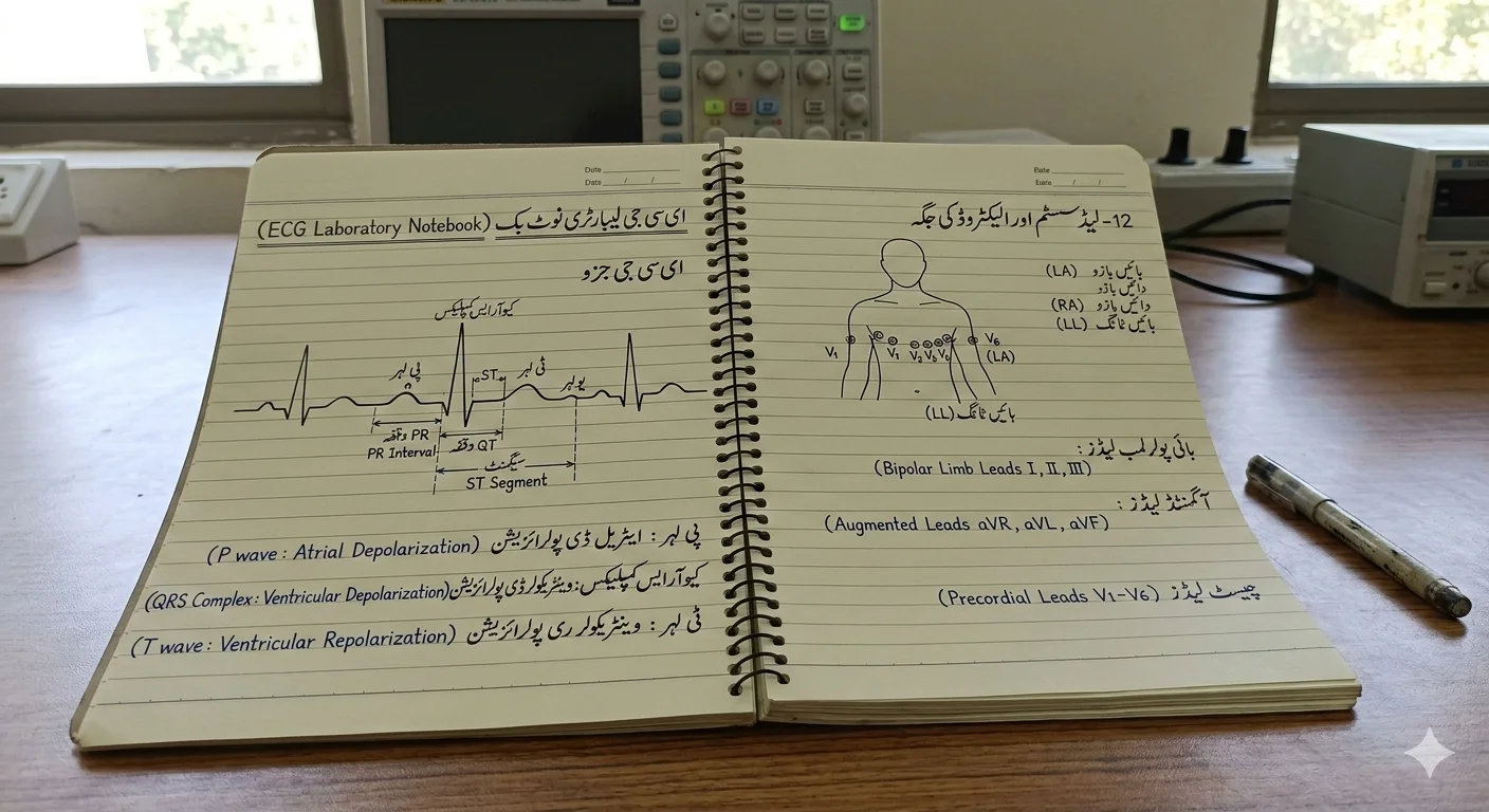
The heart operates with a rhythm that can be captured visually through ECG waveforms. Each waveform represents an electrical event during the cardiac cycle.
The P wave marks atrial depolarization, signaling that the upper chambers of the heart are contracting. This is followed by the QRS complex, which signifies ventricular depolarization—the moment when the lower chambers pump blood to the lungs and body.
Afterward comes the T wave, reflecting ventricular repolarization as muscles prepare for another contraction. Understanding these basic components is crucial for interpreting heart health.
Anomalies in these waves can indicate various cardiac issues. Recognizing what each part signifies enhances your ability to analyze rhythms effectively and diagnose conditions accurately. Embracing this knowledge lays a strong foundation for working within your ECG components lab notebook.
Types of ECG Leads
ECG leads are essential for capturing the heart’s electrical activity. They come in various types, each serving a unique purpose.
The standard limb leads include Lead I, II, and III. These provide a view of the heart’s electrical signals from different angles. They help detect arrhythmias and other abnormalities effectively.


Chest leads, or precordial leads (V1-V6), offer detailed insights into specific areas of the heart. Placed across the chest wall, they create a three-dimensional picture of cardiac function.
Augmented limb leads—AVR, AVL, and AVF—enhance the standard views by providing additional perspectives from different axes. This is crucial for comprehensive analysis.
Understanding these lead types ensures accurate monitoring during ECG procedures. Proper application aids in diagnosing conditions early on while reducing errors in interpretation.
The Importance of Proper Lead Placement
Proper lead placement is crucial for obtaining accurate ECG readings. Even slight misplacement can distort the cardiac waveforms and lead to misdiagnosis.
Each lead has a specific anatomical location that reflects different aspects of heart activity. Correct positioning ensures that you capture the electrical signals from various angles, allowing for a comprehensive view of cardiac function.
For instance, placing leads too high or low can result in misleading information about atrial or ventricular activities. This might mask underlying conditions or create false positives in your analysis.
Practicing consistent lead placement techniques helps build reliability in your recordings. It also enhances your ability to identify abnormalities with greater precision, further improving patient outcomes.
Training and routine checks on lead placements are vital practices within any ECG procedure. By prioritizing accuracy here, you’re setting a solid foundation for interpreting results effectively.
Interpreting Ecg Components Lab Notebook
Interpreting the ECG components in your lab notebook is a skill that develops with practice. Each waveform tells a story about the heart’s electrical activity. It’s essential to familiarize yourself with these patterns.
P waves indicate atrial depolarization, while QRS complexes represent ventricular depolarization. The T wave signifies repolarization of the ventricles. Recognizing these key elements allows for better understanding and analysis.
Pay attention to intervals and segments too. The PR interval can indicate conduction delays, while ST segment changes might suggest ischemia or other cardiac events.
Take notes as you study each ECG reading in your lab notebook. Document observations meticulously; they will serve as valuable references later on when reviewing cases or spotting trends in cardiac health.
Practice makes perfect—regularly interpreting various ECGs enhances both confidence and accuracy over time.
Common Abnormalities and Their Significance
Common abnormalities in ECG readings can reveal critical insights into cardiac health. Each waveform anomaly serves as a potential indicator of an underlying issue.
For instance, atrial fibrillation is marked by irregularly spaced QRS complexes and the absence of distinct P waves. This condition may lead to stroke or heart failure if left untreated.
Another common abnormality is ST-segment elevation, often signaling myocardial infarction. Identifying this promptly can save lives through immediate intervention.
Additionally, prolonged QT intervals are associated with the risk of life-threatening arrhythmias. Monitoring these changes helps clinicians act swiftly.
Recognizing these abnormalities not only aids diagnosis but also enhances treatment strategies tailored to individual patient needs. In a lab notebook setting, documenting such findings accurately allows for better trends analysis over time.
Tips for Accurate Recording in a Lab Notebook
Accurate recording in an ECG components lab notebook is essential for reliable data analysis. Start by using a consistent format. This helps maintain clarity throughout your entries.
Label each section clearly, including dates and patient identifiers. Clear headings make it easier to navigate through the information later on.
Always write legibly, whether you’re noting abnormalities or standard waveforms. If digital recordings are used, ensure that all printed outputs are dated and attached securely.
Use color coding for different types of waves or abnormalities to enhance visual understanding. This method can simplify complex interpretations during review sessions.
Don’t forget to include observational notes about the patient’s condition when the ECG was taken; these details often provide context that numbers alone cannot convey.
Regularly check your entries for completeness before moving on to new recordings, as accuracy at this stage leads to more effective analyses down the line.
Case Studies and Practice Exercises
Exploring case studies enhances your understanding of the ECG components lab notebook. Real-life scenarios allow you to apply theoretical knowledge in a practical context.
Consider a patient with chest pain. Analyzing their ECG can unveil critical insights into potential ischemic changes or arrhythmias. By reviewing such cases, you sharpen your diagnostic skills and build confidence.
Practice exercises are equally essential. Create sample ECG strips based on varying conditions like atrial fibrillation or ventricular tachycardia. This hands-on approach reinforces recognition patterns and deepens comprehension of waveforms.
Additionally, participate in group discussions about these cases. Engaging with peers encourages diverse perspectives and promotes collaborative learning.
Combining case studies with practice exercises forms a robust foundation for mastering interpretation skills within the ECG components lab notebook framework.
Conclusion
Mastering the ECG components lab notebook is an essential skill for anyone involved in cardiac care. Understanding the fundamental cardiac waveforms lays the foundation for effective interpretation and analysis. Familiarity with different types of ECG leads and their proper placement can significantly enhance your diagnostic accuracy.
Interpreting records from the ECG components lab notebook requires attention to detail, as does recognizing common abnormalities that can have significant implications for patient health. By honing these skills, you not only improve your ability to provide accurate assessments but also contribute positively to patient outcomes.
Keeping detailed notes in your lab notebook ensures a reliable reference point for future cases or when collaborating with colleagues. Engaging with case studies and practice exercises reinforces learning and builds confidence in real-world applications.
With dedication and continual practice, mastering this vital aspect of cardiac evaluation becomes achievable, empowering you to make informed decisions based on clear data insights. This journey toward proficiency enhances both individual expertise and overall healthcare quality within cardiology settings.